Overview
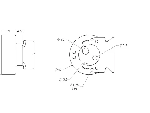
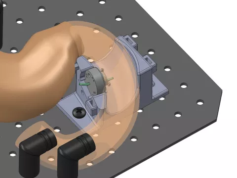
The model consists of three main components: a reusable silicone rubber stomach and duodenum (The Chamberlain Group, Great Barrington, MA), a 3D printed ampullary mount to which the chicken heart is attached, and a 3D printed base to stabilize the duodenum and to act as a guide to allow repeated insertion and extraction of the mount. The base allows the simulated ampulla to enter the duodenum perpendicular to the wall of the model through a 14mm opening. The mount has a 2.5mm pancreatic duct and a 4mm bile duct. The model allows for successful completion of ampullectomy procedural steps, including passing an ERCP scope through the pylorus to the ampulla, identification of the ampulla, positioning the snare over the ampulla, performing snare resection, specimen retrieval, pancreatic duct cannulation, and pancreatic stent insertion.

Development of an Educational Training Device
As part of a larger AdventHealth Endoscopic Resection Conference, there was a need for an Ampullectomy Training Model. Endoscopic ampullectomy is a technically challenging, high-risk procedure with limited training opportunities. A model did not exist, so we set out to develop our own. ISA worked with the Center for Interventional Endoscopy (CIE) to design and develop a training device used to improve technical skills, knowledge and confidence in performing endoscopic ampullectomy.
Dimensions, CAD Setup, Assembly, Assembly - Stent
This slideshow displays one large image or video at a time. Use the Previous and Next buttons to move between images, or use the following thumbnails slideshow to select a specific image or video to display here.
This slideshow contains a row of small thumbnails. Selecting a thumbnail will change the main image in the slideshow that is above it. Use the Previous and Next buttons to cycle through all the thumbnails, use Enter to select.


