- AdventHealth
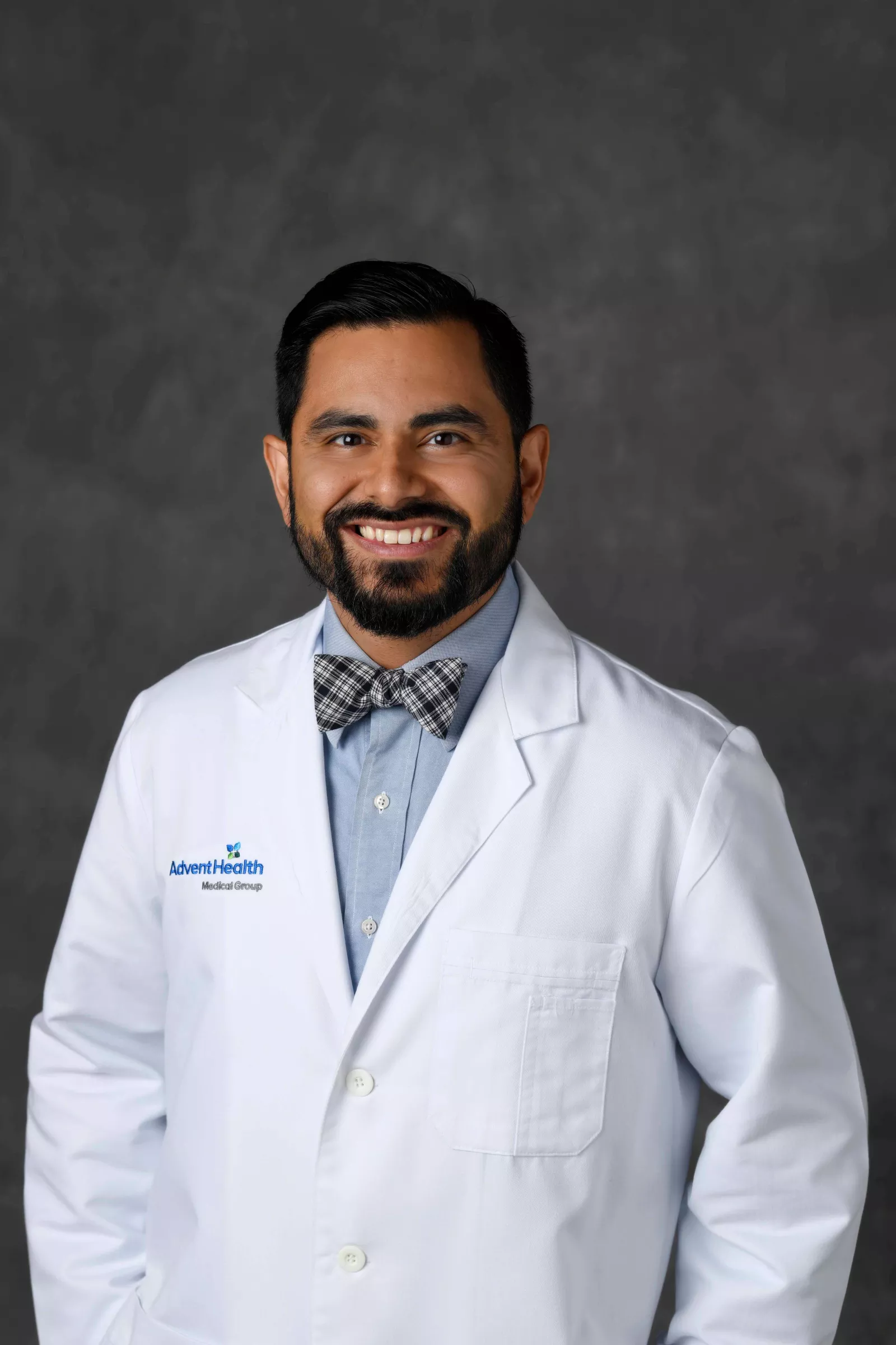
This Clinician's View is written by Joseph Lopez, MD, MBA, FAAP, AdventHealth for Children medical director for pediatric head & neck surgery.

By age 3, Paisley Battles had experienced more medical tests than many people do in a lifetime, yet she was still suffering from frequent severe headaches, dizziness, constipation, daily pain in her lower extremities and delayed speech with no clear diagnosis. One day, after Paisley experienced a syncope episode in her car seat, her parents drove straight from their home in Edgewater, Florida, to the AdventHealth for Children emergency room where the medical team admitted her. A magnetic resonance imaging (MRI) scan revealed the diagnosis — Chiari malformation type 1 (CM1).
Chiari malformations (CM) are caused by problems in the structure of the brain and skull. Affecting approximately 1 in 1,000 children, Chiari malformation type 1 is the most common form and occurs when the cerebellar tonsils (the lower part of the cerebellum) push into the foramen magnum (the opening between the skull and spinal cord). The condition develops as the skull and brain are growing and can put pressure on the brain, blocking the flow of cerebrospinal fluid.
At AdventHealth for Children, James Baumgartner, MD, pediatric neurosurgeon, and I recently began taking a new surgical approach to treating Chiari malformation type 1 with the aim of improving patient outcomes: suboccipital posterior vault distraction osteogenesis (PVDO). On July 19, 2023, Paisley was our first case, and we have since successfully performed 4 additional suboccipital PVDO procedures.
The Challenges of Chiari Malformation Type 1
While Chiari malformation type 1 can be asymptomatic, the altered cerebrospinal fluid dynamics along with the pressure from the cerebellum on the spinal cord or lower brainstem can cause troubling symptoms to arise in some patients. The most common include:
- Headaches (especially after coughing, sneezing or straining)
- Neck pain
- Dysphagia (difficulty swallowing)
- Problems with balance and coordination
- Numbness and tingling of the hands and feet
- Vertigo (dizziness)
- Dysarthria (slow or slurred speech) and hoarseness
- Ocular disturbances
- Sleep apnea
As in Paisley’s case, when patients present with symptoms, we use MRI to help make the proper diagnosis. The presence of associated cranial nerve deficits, brainstem dysfunction, kyphosis (excessive forward rounding of the upper back), and syringomyelia (the development of a fluid-filled cyst within the spinal cord), particularly in young patients in early stages of disease, are indications for operative intervention. Paisley’s MRI revealed crowding of the foramen magnum and a mass effect on her cervicomedullary junction, the region where the brainstem continues as the spinal cord, indicating a potential benefit from surgical treatment.
Traditional Surgical Approach to Treating Chiari Malformation Type 1
For the past 40 years, suboccipital posterior fossa decompression (PFD) has served as the standard surgical approach to treating Chiari malformation type 1. It involves removal of a portion of the skull to create more space for the cerebellum. PFD is typically performed alone or with associated duraplasty, which involves opening the dura, the thick membrane covering the brain and spinal cord, and sewing a patch to enlarge it, further relieving pressure. Atlas laminectomy, the removal of part of the vertebra to create more space, is also frequently performed with PFD.
While typically effective, this approach comes with risks and challenges. It involves removing bone, which artificially creates space, possible cervical instability and cerebrospinal fluid leakage (pseudomeningocele). With PFD, there is also a risk of stroke, resulting in damage to the cerebellum.
Applying an Effective Craniosynostosis Surgical Approach to Treating Chiari Malformation Type 1
Posterior cranial vault distraction osteogenesis (PVDO) is utilized routinely for treatment of multi-suture craniosynostosis, a rare birth defect that occurs when one or more joints in a baby’s skull fuse together before the brain finishes developing. The procedure is designed to expand the back of the skull, giving the brain room to grow.
Chiari malformation type 1 frequently occurs with craniosynostosis, and surgeons have found that performing PVDO improves symptoms of both conditions. That is why after careful research and consideration, we have started using a suboccipital PVDO surgical approach to treat appropriate patients who suffer only from Chiari malformation type 1 without craniosynostosis. We believe suboccipital PVDO offers a safe option for these patients and can enhance their outcomes. Compared to PFD, PVDO creates space but without having to cut into the dura or remove bone. In fact, PVDO promotes the formation of new bone (ossification) in the posterior fossa, allowing the cerebellum to unherniate itself, thus creating more space as the skull grows.
Current published literature on the use of PVDO to treat Chiari malformation type 1 is limited to case reports and small retrospective case series. In 2022, one such report presented a case series of 9 patients with Chiari I malformations treated with distraction osteogenesis, along with a novel technique to safely and effectively expand the posterior fossa while minimizing the risk of cerebellar ptosis. All patients treated experienced improvements in prior presenting symptoms.
How Suboccipital Posterior Cranial Vault Distraction Osteogenesis is Performed
When using suboccipital PVDO to treat patients with Chiari malformation type 1, the main goal is to create more space in the posterior fossa, alleviating the compression and pressure on the cerebral tonsils and brainstem. Secondarily, gradual distraction facilitates new bony growth, which helps maintain the additional space posteriorly in the long-term.
While we continue to evaluate each case and enhance our approach to optimize treatment, the basic suboccipital PVDO procedure on patients with Chiari malformation type 1 involves several key components:
- Our team uses customized, computer-assisted guides to plan each patient’s procedure, therefore personalizing the surgery to each patient’s anatomy.
- The incision is hidden in the hairline around the back of the child’s skull bone.
- Small metal distractors are attached across the incision at preplanned vectors. Placed under the skin, the distractors have posts that extend through the skin.
- The extenders allow for post-surgical daily adjustment of the distractors over the course of 2-3 weeks, which slowly and painlessly stretches the two pieces of bone apart at a rate of 1 millimeter a day (distraction osteogenesis).
- Once the bones are determined to be in the proper position, the distractor adjustment is halted.
- Over a period of approximately 2-3 months, new bone grows to fill the gap.
- A second surgical procedure is performed to remove the distractors after the new bone has healed.
Effective provider and caregiver education on post-operative care is critical to alleviating complications and re-operation rates. For Chiari malformation type 1 patients, the suboccipital PVDO approach is only appropriate if they have not previously had PFD. However, having PVDO does not preclude a patient from having PFD later if determined necessary. While PVDO does involve a longer incision, our team works to ensure the scar is imperceptible, and hair follicles eventually grow through the scar.
Improving Patient Outcomes and Helping Children Feel Whole
At AdventHealth for Children, we believe in taking a whole-person approach to the care we provide, and our goal is to help every child return to just being a child. I’m pleased to report that of the 8 suboccipital PVDO surgeries we have performed to date on children with Chiari malformation type 1, all have experienced complete resolution of their symptoms. For children like Paisley, this can be life-changing.
As soon as the morning after her surgery, Paisley was talking in full sentences, something her parents Krysta and Justin said had not happened before. Paisley’s debilitating headaches, leg pain, neuropathy and incontinence have also ceased. Now 4 years old, she has no limitations on her physical activity and just started pre-K where her parents report she is performing at the top of her class in almost all subjects. Paisley loves to dance and is working on perfecting her cartwheel. She also enjoys playing soccer with her siblings and hopes to start karate soon.
Optimizing Care for Patients with Chiari Malformation Type 1
Our team remains committed to innovation that enhances care and optimizes outcomes for our patients. With every suboccipital PVDO case we perform, we are closely evaluating each step of the process to identify opportunities to refine our approach. To that end, we are currently working to develop a new distractor device that we believe will better meet the specific needs of performing suboccipital PVDO on patients with Chiari malformation type 1.
Additionally, we recently conducted a systematic review of the published literature for PVDO as a treatment for Chiari malformation type 1, and our work has been accepted for publication in an upcoming issue of the Journal of Craniofacial Surgery. Looking ahead, we also hope to collaborate with other high-volume craniofacial centers on additional research efforts and to further refine techniques and protocols that will continue to transform the treatment of patients with Chiari malformation type 1, alleviating their symptoms and helping them to grow up healthy, happy and whole.


